The Pupillary Light Reflex
A minority of optic tract fibers bypass the LGN entirely, traveling dirctly to the midbrain. One of their targets in the midbrain is the pretectal area, which mediates the pupillary light reflex. This reflex can be demonstrated by shining a light in one eye; if all is working correctly, both pupils will constrict.
The optic nerve is responsible for the afferent limb of the pupillary reflex and the oculomotor nerve is responsible for the efferent limb of the pupillary reflex.
The pupillary reflex pathway begins with retinal ganglion cells, which convey information from photoreceptors to the optic nerve (via the optic disc)
The optic nerve connects to the pretectal nucleus of the upper midbrain, bypassing the lateral geniculate nucleus and the primary visual cortex.
The posterior commissure interconnects the pretectal nuclei, mediating the consensual pupillary light reflex.
From the pretectal nucleus, axons connect to neurons in the Edinger-Westphal nucleus, whose axons run along both the left and right oculomotor nerves.
Oculomotor nerve axons synapse on ciliary ganglion neurons.
Fibres from the ciliary ganglion innervates the constrictor muscle of the iris
Schematic representation of the visual pathway
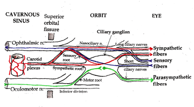
Pathways in the Ciliary ganglion. Green = parasympathetic; Red = sympathetic; Blue = sensory
.