Relative afferent pupillary defect (RAPD)
Assessment of RAPD (Relative afferent pupillary defect)
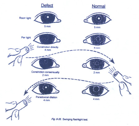
For assessing the indirect response, utilize the swinging flashlight test by Placing the penlight in front of the right eye (OD) The examiner initially watches the Right eye for a direct response and the left eye for a consensual response. Both pupils should constrict The light should then be swung to the left eye (OS). The normal pupil will constrict to the direct light as the light swings back and forth, and the normal pupil will also constrict to indirect light, having a consensual response. RAPD constricts to consensual light and not to direct light. Next repeat the above starting with the left eye and swinging to the right eye.

Abnormalities of the Pupil and Their Characteristics Relative Afferent Pupillary Defect (RAPD) – also referred to as APD and Marcus-Gunn Pupil
Objective sign of an asymmetrical lesion of the anterior visual pathway (retina, optic nerve, chiasm or optic tract)
Seen with major retinal lesions or neurological lesions of the anterior visual pathway.
The presence of a RAPD and the absence of gross ocular disease indicate a neurological lesion in the anterior visual pathway and the importance of this physical sign cannot be over emphasized.
Grading Scale:
-
RAPD Grade 1+: A weak initial pupillary constriction followed by greater redilation
-
Grade 2+: An initial pupillary stall followed by greater redilation
-
Grade 3+: An immediate pupillary dilation
-
Grade 4+: No reaction to light – Amaurotic pupil
Conditions associated with an RAPD
A) Unilateral optic nerve diseases:
-
i) Anterior ischemic optic neuropathy: ii) Arteritic (Giant Cell Arteritis) and Non-arteritic iii) Optic neuritis iv) Compressive optic neuropathy: a) Optic nerve tumor b) Traumatic optic neuropathy c) Asymmetric glaucoma d) Homonymous hemianopsia (Lesions involving pupillary pathway)
B) Retinal conditions:
-
i) Central retinal arterial occlusion ii) Branch retinal arterial occlusion iii) Ischemic central retinal vein occlusion iv) Retinal detachment, greater RAPD if involving macula
